E. Ersoy,1 M. Ozdogan,1 H. Kulaçoglu1 and H. Gundogdu1
1Department of General Surgery, Ankara Ataturk Research and Educational Hospital, Ankara, Turkey (Correspondence to E. Ersoy:
Received: 29/01/06; accepted: 23/05/06
EMHJ, 2008, 14(5): 1222-1224
Introduction
Retained postoperative foreign bodies, of which sponges are the most common, are a rare situation. This may be due to the care of the surgical team or a reluctance to publish incidences that could lead to medicolegal problems. Despite this rarity in reporting, retained foreign bodies appear to be encountered more commonly than expected. All surgical tools used during an operation can become a retained foreign body. Surgical instruments, sponges and drains left in the operation site may be responsible for bizarre and varied complications. We describe a case of a Penrose drain retained in the axillary region discovered during the dissection of the lymph nodes due to epidermoid carcinoma.
Case report
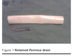
A 50-year-old man presented to our clinic in December 2005 with a recurrent mass in the left axillary region. He had suffered a burn 15 years before. Ten months before presenting to us, he was found to have a Marjolin ulcer on the burn scar indicating epidermoid carcinoma. He was admitted to another hospital where radical excision of the tumour was performed together with lymph node dissection, which on pathology examination confirmed metastasis. He then underwent radiotherapy of the lymph node metastasis. He was admitted to our clinic 10 months later with a mass in the ipsilateral axillary region which had appeared about 3 weeks before. Physical examination revealed a mass of 3 × 4 cm in the left axillary region with no inflammation or cellulites on the skin. There was a burn scar on the left hand and forearm. Local ultrasound scan also showed a mass of 4 cm diameter. We decided to excise the mass assuming that it was a metastatic focus. During the operation, we found a cavity surrounded by a pseudocapsule which contained a Penrose drain of 10 cm length (Figure 1). The foreign body was excised with the surrounding tissue and pathological examination revealed an epidermoid carcinoma in the pseudocapsule tissue. The patient is still being followed up.

Discussion
The actual incidence of retained foreign bodies is difficult to estimate but has been reported to be 1 in every 3000 procedures [1]. This inadvertent complication most frequently occurs in gynaecological and upper abdominal surgical procedures and generally after emergency operations [2]. Gossypibomas, retained surgical sponges, are the most commonly seen retained foreign bodies.
Retained foreign materials may lead to serious complications, particularly when found to be intra-abdominal, such as perforation, obstruction, fistula formation, sepsis or even death. On the other hand, sometimes they can stay undetected without causing any symptoms for months or years. There are reports of retained foreign bodies remaining undetected for 40 years between the onset and diagnosis [3,4]. In our case, the Penrose drain led to a pseudo-tumour reaction after 10 months but at the same time within the tumour, epidermoid cancer was found and histopathologically confirmed. Therefore it is possible that the axillary mass was a cancer metastasis and the drain was discovered coincidentally.
Ultrasound can be used to investigate and a well delineated mass containing wavy internal echo with a hypoechoic rim and a strong posterior acoustic shadowing should suggest the possibility of foreign body retention [5]. Our case showed a hyperechoic solid mass but the drain was not visible. Probably the degenerated and healed tissue resulting from the radiotherapy concealed the drain.
Although computerized tomography and ultrasonography are widely used to detect a retained foreign body, 30% are in fact found during an operation [6], as in our case. Early identification of a retained foreign body is important, otherwise it may lead to complications and unnecessary invasive procedures and operations [7].
In our case, it is unclear how the Penrose drain was left in the axillary region; whether it was forgotten or broken while being removed but both situations present a medicolegal problem. There are some cases in the literature similar to ours but most of them are about foreign bodies left in the surgical cavity of mastectomies [8,9].
In general, closely following operating room and general surgery clinic procedures (adequacy of operative notes, quality of postoperative care and follow-up, state of discharge summaries, clinical and significant event audit, etc.) should be enough to prevent the problems of retained foreign bodies, but nonetheless they still occur. Some authors suggest routine radiography after surgery but this does not seem to be cost-effective. It is reported that false negative rates in detecting retained sponges using radiography varied between 3% and 25% depending on the type of sponge [10]. Although retention of foreign bodies after surgery is rare, it raises worrying issues of patient safety and surgeon/nurse responsibility. Every effort must be made to prevent its occurrence to avoid unnecessary morbidity. Mechanisms to do this include.
- Continuous medical training to ensure strict adherence to surgical rules.
- Use of sponges or instruments with radio-opaque markers.
- Use of drain fixing sutures.
- Sponge and instrument counting before the procedure ends.
- Performance of routine postoperative wound and cavity exploration before wound closure.
- Avoidance of shifting during the operation where possible.
- Effective follow-up after surgery.
References
- Jarbou SM, Al-Kurdi M, Al-Daod K. Pseudotumour due to retained surgical sponge (gossypiboma). Eastern Mediterranean health journal, 2004, 10(3):455–7.
- Bani-Hani KE, Gharaibeh KA, Yaghan RJ. Retained surgical sponges (gossypiboma). Asian journal of surgery, 2005, 28(2):109–15.
- Liessi G et al. Retained surgical gauzes: Acute and chronic CT and US findings. European journal of radiology, 1989, 9(3):182–6.
- Sakayama K et al. A 40 year old gossypiboma (foreign body granuloma) mimicking a malignant femoral surface tumor. Skeletal radiology, 2005, 34(4):221–4.
- Chau WK, Lai KH, Lo KJ. Sonographic findings of intraabdominal foreign bodies due to retained gauze. Gastrointestinal radiology, 1984, 9:61–3.
- Yildirim S et al. Retained surgical sponge (gossypiboma) after intra-abdominal or retroperitoneal surgery: 14 cases treated at a single center. Langenbecks archives of surgery, 2006, 391(4):390–5.
- Williams RG, Bragg DG, Nelson JA. Gossypiboma – the problem of the retained surgical sponge. Radiology, 1978, 129(2):323–6.
- Karcaaltincaba M et al. Breast abscess mimicking malignant mass due to retained Penrose drain: diagnosis by mammography and MRI. Clinical imaging, 2004 28(4):278–9.
- De Souza GA. Penrose drain as a foreign body in the breast. The breast journal, 1999, 5(3):208–10.
- Revesz G, Siddiqi TS, Buchheit WA. Detection of retained surgical sponges. Radiology, 1983, 149:411–3.


